
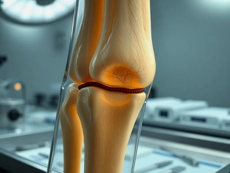
Bone shape regenerates within three weeks while material strength continues to mature
Researchers at Texas A&M tracked regeneration in mouse digit tips and found a clear temporal gap between morphological reconstruction and mechanical maturation. Using a fingertip-amputation model, they observed that the bone’s gross geometry and continuity typically reappear by about day 21 after injury, but nanoscale mechanical properties keep evolving through days 28–31. This separation means that a healed-looking bone can still be mechanically immature.
How the experiment measured structure and mechanics across scales
The team combined three complementary methods to capture both visible anatomy and microscale mechanics. First, micro-computed tomography (micro-CT) produced high-resolution 3D images to quantify bone volume, cortical geometry, and gross morphological closure over time. Second, atomic force microscopy (AFM) mapped local mechanical properties—such as indentation modulus and surface stiffness—at the micron-to-submicron scale across regenerating regions. Third, finite element modeling (FEM) took micro-CT geometries and AFM-derived material properties to predict stress and strain distributions in the regenerating digit under physiological loading.
Why morphology and mechanics follow different timelines
Morphological reconstruction depends primarily on cellular patterning and matrix deposition by blastema-derived cells that repopulate the defect and reestablish anatomical continuity. These processes—cell migration, proliferation, and bulk mineral deposition—can restore shape relatively quickly. Mechanical maturation, however, requires hierarchical remodeling: mineral crystal growth, collagen crosslinking, and microstructural organization that tune local stiffness and toughness. Those nanoscale and microscale processes proceed slower, which the AFM measurements captured as continuing increases in local stiffness after the overall shape had formed.
What the finite element models revealed about load distribution
Integrating AFM-derived stiffness maps into FEM showed that early in regeneration, mechanical loads concentrate unevenly across anterior and posterior surfaces and across the regenerating interface. Over time, as local stiffness increases and the geometry normalizes, the predicted load distribution becomes more uniform. The models therefore illustrate how delayed mechanical maturation can cause transient stress concentrations that might influence remodeling pathways or susceptibility to reinjury under premature loading.
Practical implications for rehabilitation and loading
The study does not endorse specific clinical protocols for humans, but it highlights a general principle: restoring external anatomy does not guarantee full mechanical competence. Decisions about when to permit weight-bearing, progressive loading, or return to activity should consider the potential lag in material-level recovery. That assessment ideally uses objective measures (imaging biomarkers, patient symptoms, functional testing) and clinical context—fracture type, fixation stability, comorbidities—rather than visual union alone.
Evidence that neighboring bone adapts during repair
AFM and imaging also detected alterations in adjacent, non-amputated bone tissue. Nearby regions showed changes in material properties and microarchitecture consistent with a coordinated, multicompartment repair response. These changes suggest paracrine signaling, altered mechanical environment, or remodeling stimuli extend beyond the primary injury site, contributing to whole-digital adaptation.
Relevance to genetic models and Down syndrome research
Investigators linked to Dr. Lindsay Dawson are expanding these multiscale methods into genetic models, including Down syndrome, to examine how genotype and age shape regenerative capacity and material recovery. Using AFM and micro-CT in tandem provides a sensitive approach to detect genotype-dependent delays in mechanical maturation that would be missed by morphology-only readouts.
Limitations and what the findings do not prove for humans
Key limitations constrain direct translation. The work uses a mouse digit-tip model, which regenerates differently than most human bones. Microenvironmental cues, scale-dependent mechanics, and healing timelines differ between species and skeletal sites. These results do not establish that human fractures reach full mechanical competence by a fixed day count nor that earlier loading will harm or help in specific clinical scenarios without controlled trials.
Experimental timeline and concrete results summary
– Morphology: micro-CT showed restoration of gross bone continuity and shape by ~21 days post-injury in the mouse digit-tip model.
– Local mechanics: AFM revealed progressive increases in indentation modulus and local stiffness that continued through days 28–31.
– Mechanics-to-structure integration: FEM predicted initially uneven stress patterns that normalized as microscale stiffness matured.
How researchers can build on these findings—step-by-step suggestions
1. Replicate across skeletal sites: apply AFM + micro-CT + FEM to other mouse bones (eg, long bones) to test whether the morphology-mechanics lag is general.
2. Time-course densification: sample additional intermediate time points (eg, days 14, 17, 24) to resolve exact onset and trajectory of mechanical maturation.
3. Functional loading studies: implement controlled, progressive mechanical loading in vivo while monitoring AFM properties and micro-CT geometry to establish causality between load and maturation rate.
4. Genetic and age comparisons: test models with known bone-healing deficits (aging, Down syndrome, osteoporosis models) to quantify differences in mechanical maturation timelines.
5. Translate candidate biomarkers: evaluate noninvasive imaging or serum markers correlated with AFM-derived stiffness to enable clinical monitoring strategies without nanoscale probes.
Takeaway for researchers and discharge
The central takeaway: anatomical union and visible bone shape reappearance occur earlier than complete mechanical restoration at the tissue level. Multiscale assessment—combining imaging, nanoscale mechanical mapping, and computational modeling—reveals a staggered recovery that could inform experimental design, timing for functional tests, and hypotheses about when mechanical stimuli most effectively promote durable repair.
Be the first to comment